Understanding Skull Base Anatomy: Designs and Works
The skull base fills in as the underpinning of the skull, supporting and safeguarding vital designs while interfacing with the cranial and facial districts. This unpredictable physical region holds huge significance because of its association with different tactile, apprehensive, and vascular frameworks.
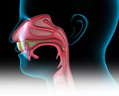
Bones and Designs of the Skull Base
The skull base contains a few bones, including the front-facing, ethmoid, sphenoid, occipital, and fleeting bones. These bones structure complex shapes that make different openings and waterways, lodging critical designs like the mind, cranial nerves, veins, and spinal lines. For example, the ethmoid bone adds to the nasal cavity and structures a part of the orbit.
Divisions and Importance
The skull base is partitioned into three particular areas – the front, center, and back fossae, and houses vital cerebrum structures. The foremost fossa made fundamentally out of the front-facing bone, safeguards the cerebrums. Conversely, the center fossa, shaped by the worldly and sphenoid bones, covers pivotal regions like the fleeting curves and pituitary organs. The back fossa, mostly comprised of the occipital bone, incorporates the brainstem, cerebellum, and fundamental cranial nerves, directing coordination and vital flagging.
Understanding skull base anatomy is crucial in clinical strengths like neurosurgery, otolaryngology (ENT), and radiology. Specialists and experts require complex information to carry out exact methods, like growth evacuation and fixing cerebrospinal liquid leaks. ENT experts frequently oversee conditions like sinus problems and nasal section cancers.
Clinical Applications and Imaging Procedures
Headways in imaging advances, similar to X-ray (Attractive Reverberation Imaging) and CT (Registered Tomography) checks, have fundamentally helped with picturing the skull base. These imaging methods give nitty gritty perspectives on bone designs, delicate tissues, and abnormalities within the skull base, supporting conclusions and treatment anticipating different pathologies.
Future Points of View and Exploration Regions
Continuous exploration in skull base anatomy centers around working on careful methods, investigating negligibly obtrusive methodologies, and propelling imaging modalities. Understanding the complicated connections between structures in this area keeps on preparing for creative medicines and upgraded patient results.
Clinical Importance and Careful Contemplations
Skull base anatomy holds huge clinical pertinence, particularly in neurosurgery. Methods including the evacuation of cancers or tending to abnormalities in this locale request accuracy and ability because of the proximity of critical designs. Specialists exploring the complexities of the skull base require a profound comprehension of its anatomy to limit possible harm to vital nerves, veins, and the cerebrum. Additionally, headways in careful methods, like endoscopic methodologies, have altered the treatment of skull base pathologies, offering negligibly obtrusive choices with decreased recuperation times for patients.
Pathologies and Problems
A scope of pathologies and problems can influence the skull base, causing huge clinical difficulties. Growths, both harmless and dangerous, maybe fostered around here, influencing different capabilities and presenting dangers to neighboring designs. Conditions like meningiomas, chordomas, and pituitary adenomas normally include the skull base. Also, wounds bringing about breaks or injury to this area can prompt difficulties, including cerebrospinal liquid leaks, which necessitate brief clinical intercession and specific consideration.
Arising Advancements and Treatment Modalities
Headways in innovation keep on altering the conclusion and treatment of skull base problems. Advancements in picture-directed careful route frameworks consider exact and designated mediations, diminishing usable dangers and upgrading results. Besides, an examination into customized medication and sub-atomic focusing offers promising roads for custom-made treatments in treating explicit skull base pathologies. Novel treatment modalities like proton pillar treatment and immunotherapy are additionally acquiring consideration for their true capacity in overseeing skull base growths and related conditions.
Conclusion
The complexities of skull base anatomy highlight its vital job in keeping up with neurological and tactile capabilities while filling in as a foundation for different clinical mediations. Its multi-layered nature requires a far-reaching understanding across clinical disciplines, underscoring its importance in both clinical practice and continuous exploration tries.
(FAQs):
1. What designs are housed within the skull base?
The skull base houses vital designs including the mind, cranial nerves, veins, and spinal line. It comprises different bones framing unmistakable districts, like the foremost, center, and back fossae.
2. How is skull base anatomy important in clinical practices?
Understanding skull base anatomy is essential, especially in neurosurgery and otolaryngology (ENT), directing surgeries and treatment anticipating growths, wounds, and problems influencing this area.
3. What imaging procedures are utilized to envision the skull base?
Imaging modalities like X-ray (Attractive Reverberation Imaging) and CT (Registered Tomography) filters give itemized perspectives on the skull base, supporting diagnosing pathologies and arranging careful intercessions.